
What Is an Echocardiogram (Echo)?
An echocardiogram, often called an echo, is one of the most common and useful tests used in cardiology. It provides a clear and detailed picture of how your heart is working by using sound waves (ultrasound) to create moving images. Unlike an ECG, which measures electrical activity, an echocardiogram lets your cardiologist actually see your heart’s structure and function in real time.
Many people come to us unsure what the test involves or why it’s needed. In this guide, we’ll explain what an echo is, what it can show, when it’s recommended, and what to expect during the test.
What Is an Echocardiogram?
An echocardiogram is a non-invasive ultrasound scan of the heart. It uses high-frequency sound waves to produce live images of your heart’s chambers, valves, and surrounding blood vessels.
These images allow cardiologists to assess how well your heart is pumping blood, whether the valves are opening and closing correctly, and whether there are any signs of damage, weakness, or abnormal movement in the heart muscle. Because it gives such detailed information, an echocardiogram is one of the most valuable tools for diagnosing and monitoring a wide range of heart conditions.
When Is an Echocardiogram Needed?
An echocardiogram is usually recommended when your doctor or cardiologist suspects a structural or functional problem with the heart. It can also be used to track progress after treatment or surgery.
You may be referred for an echocardiogram if you experience:
- Chest pain or discomfort
- Shortness of breath
- Fatigue or dizziness
- Swelling in your ankles or legs
- A heart murmur (detected during examination)
- Irregular heart rhythms or palpitations
Echocardiograms are also used to monitor conditions such as:
- Heart failure – assessing how efficiently the heart is pumping blood
- Heart valve disease – checking for leaks or narrowing
- Cardiomyopathy – evaluating the size and shape of the heart
- Congenital heart defects – identifying structural abnormalities present from birth
- Coronary artery disease (CAD) – detecting areas of reduced blood flow
If you’re unsure whether you might need one, a cardiologist can assess your symptoms and decide whether an echo is the right test for you.
Types of Echocardiogram
There are several types of echocardiogram, each providing different information about your heart’s health:
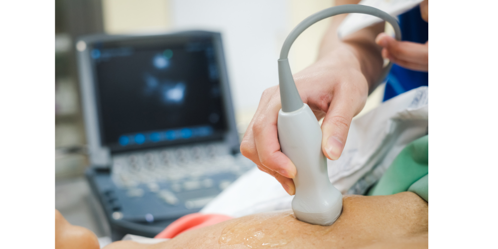
1. Transthoracic Echocardiogram (TTE)
This is the most common type. A handheld probe is placed on your chest to send and receive ultrasound waves, which are used to create moving images of your heart.
2. Transoesophageal Echocardiogram (TOE)
In some cases, clearer images are needed. A thin probe is passed gently down the oesophagus (food pipe), which sits close to the heart, providing high-quality images of its structure.
3. Stress Echocardiogram
This test evaluates how your heart performs under physical stress, such as light exercise or medication that mimics activity. It’s often used to detect coronary artery disease.
4. Doppler Echocardiogram
This variation measures the direction and speed of blood flow through your heart and valves, helping to detect abnormalities like regurgitation (leakage).
Your cardiologist will recommend the most suitable type depending on your symptoms and medical history. For a full breakdown of these tests, the British Heart Foundation offers a detailed guide on echocardiograms.
How to Prepare for an Echocardiogram
Most echocardiograms require little to no preparation. You can usually eat and drink normally beforehand, and you won’t need to take any special medication.
However:
- For a transoesophageal echocardiogram, you’ll need to avoid food and drink for a few hours before the test.
- If you’re having a stress echocardiogram, wear comfortable clothes and shoes suitable for light exercise.
Your cardiologist will provide full instructions in advance of your appointment.
What Happens During the Test?
A standard echocardiogram is simple and completely painless. Here’s what to expect:
- You’ll be asked to remove clothing from your upper body and lie on an examination couch.
- Small sticky sensors (electrodes) may be attached to your chest to record your heart rate.
- A small amount of gel will be applied to your chest to help the ultrasound probe make contact.
- The sonographer or cardiologist will move the probe around your chest to capture images from different angles.
- You might be asked to change position or hold your breath briefly to get clearer pictures.
The test usually takes about 20–40 minutes. Afterwards, you can return to normal activities straight away.
What an Echocardiogram Can Show
Echocardiograms provide valuable information about:
- The size and shape of your heart
- How well your heart muscle is pumping (known as ejection fraction)
- The function of the heart valves
- Blood flow through the chambers and arteries
- Any fluid build-up around the heart
- Structural issues, such as thickened muscle walls or dilated chambers
The results help cardiologists diagnose a wide range of conditions, from heart failure and valve disease to cardiomyopathy and congenital abnormalities.
Are There Any Risks?
An echocardiogram is a safe, non-invasive test with no known side effects. It doesn’t use radiation and is suitable for people of all ages, including children and pregnant women. In the case of a transoesophageal echocardiogram, mild throat discomfort may occur briefly afterwards, but this usually passes quickly.
After the Test
Once your echocardiogram is complete, your cardiologist will review the images and explain what they show. If any abnormalities are found, further tests such as an ECG, CT coronary angiogram, or blood tests may be recommended.
Many patients find their results are completely normal, offering reassurance and peace of mind. If a problem is detected, early identification means treatment can begin quickly, helping to prevent complications later on.
Why Checking Early Matters
Heart conditions often develop gradually, so early diagnosis is key to maintaining good health. Echocardiograms allow cardiologists to detect small changes in heart function before symptoms worsen.
WKCP uses echocardiography as part of our comprehensive diagnostic service, helping patients across Kent and East Sussex understand their heart health and access treatment promptly. We believe that regular heart checks are an important part of maintaining overall wellbeing especially for those with risk factors such as high blood pressure, diabetes, or a family history of heart disease.
Summary
An echocardiogram is one of the most valuable and widely used tests in modern cardiology. It provides clear, real-time insight into how the heart is working, helping detect conditions early and guide effective treatment. Whether you’ve been referred for an echo because of symptoms or want a general heart assessment, it’s a simple, painless, and highly informative test.
Our clinic offers fast access to echocardiograms and other cardiac tests, carried out by experienced specialists in a calm and professional setting. If you’ve been advised to have an echo, or if you’re concerned about symptoms such as breathlessness, chest discomfort, or fatigue, you can contact us here to arrange a consultation. For more detailed information, visit the NHS echocardiogram guide or Mayo Clinic’s information.